
Content verified by


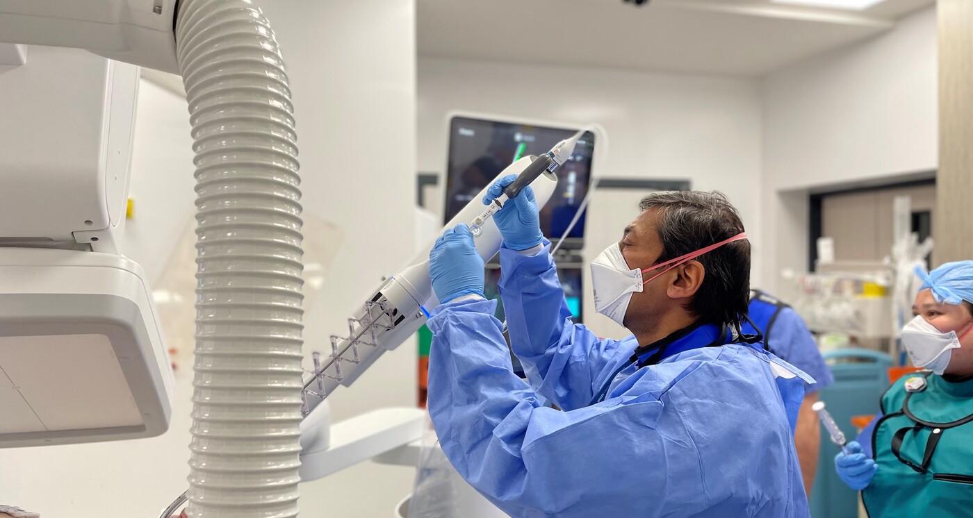
A pioneering technology, used for the first time in the UK by specialists at Royal Brompton Hospital, allows clinicians to biopsy lung tissues, including small and hard-to-reach nodules, with robotic assistance. The Ion endoluminal system (Ion) offers the potential to transform lung cancer diagnostics, through the identification of cancerous nodules at an earlier stage, when the likelihood of patient survival is greatest.
What is Ion robotic-assisted lung biopsy?
Ion (Ion endoluminal system), created by Intuitive, is a pioneering diagnostic platform that enables the robotic-assisted collection of tissue samples from almost any lung nodule. Ion procedures are minimally invasive and performed with a very thin and ultra-flexible catheter, with a camera on the end.
It is inserted via the mouth and controlled by a clinician to access the area of a patient’s lung that requires biopsy. Ion offers greater reach, flexibility and precision than traditional flexible or navigation bronchoscopic techniques.
Once the catheter is in place, multiple biopsies can be taken from an area of tissue, with a high degree of precision and accuracy. “This allows us to potentially diagnose these cancers earlier, as we can sample lung nodules of 6mm in size, or even smaller,” explains Professor Shah, “meaning we can deliver treatment promptly, reduce uncertainty, and increase patient survival rates.”
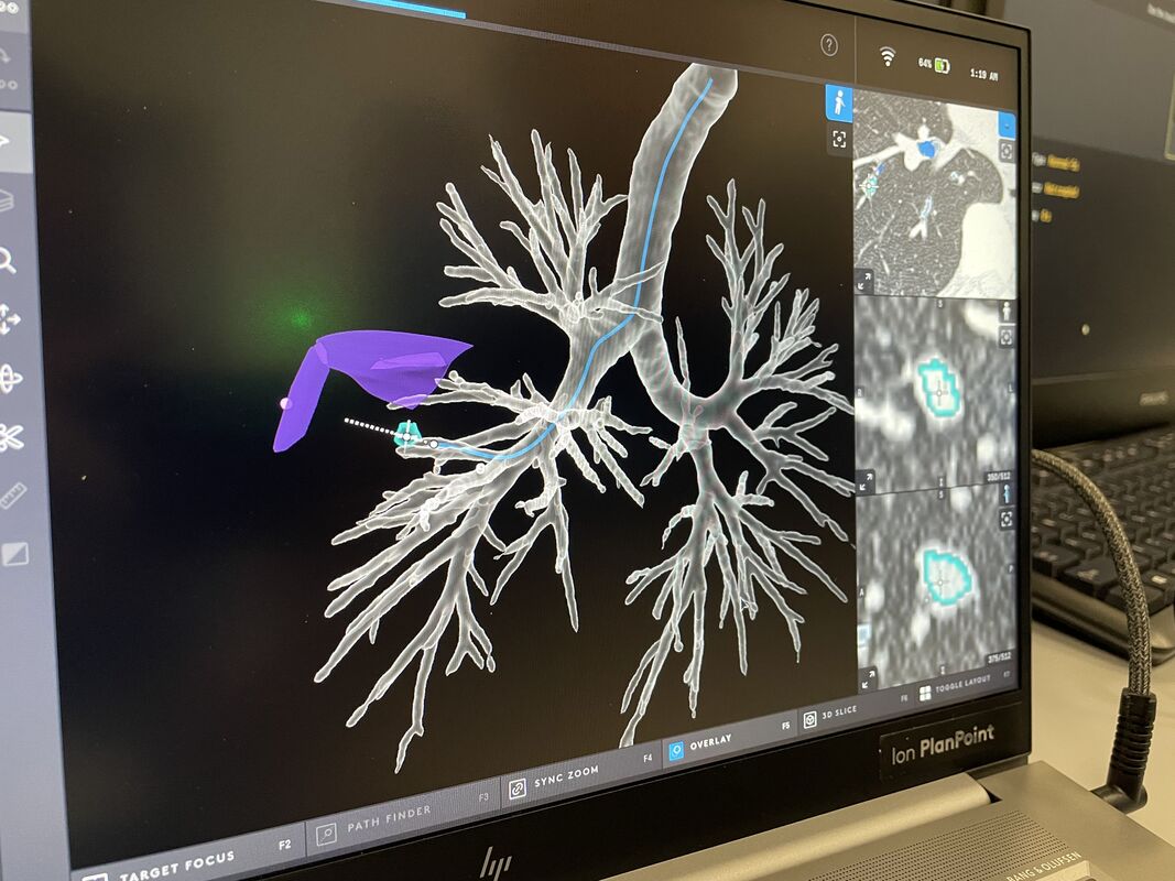
Ion 3D roadmap to guide accurate lung biopsy and surgery
Detecting lung cancer sooner
“Typically, the vast majority of our lung cancers are diagnosed when the nodule or the spot is already quite large, with cure rates between 64-70% for tumours around 30mm in size, providing there has been no other spread,” explains Professor Shah.
However, if such tumours are identified at a size smaller than 20mm, the cure rate is 83%. If a patient is diagnosed when the tumours are smaller than 10mm, the cure rate is 92%.
“As Ion can be used to biopsy very small nodules, if such a lung cancer is diagnosed, patients can begin treatment earlier, when the likelihood of survival is greatest,” explains Dr Orton.
3D roadmap for greater accuracy and safety
3D roadmap of the lungs with the proposed access routes to the nodule(s) shown by the blue line
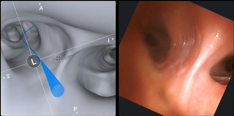
To facilitate clinicians using Ion to navigate the patient’s airways, and collect samples, the system creates a 3D roadmap of the lungs, highlighting the lung nodule(s) selected for biopsy within.
Following the collection of tissue samples, the Ion catheter can be used to place a small marker called a fiducial, to identify the area(s) of the lung needing to be removed at a subsequent surgery, allowing for even more precision.
The procedure is carried out as a day case in around 35 minutes (although it is completed in as little as 20 minutes in some cases) under general anaesthesia, with patients able to recover at the hospital for 2 hours before returning home. Some patients even return to work the day after the procedure.
Existing biopsy methods use a needle placed through the chest wall to acquire biopsies, guided by CT imaging technology for the greatest accuracy. These methods are suitable when potential tumours are larger, and have a diagnostic accuracy rate of 95.3% at our hospitals. Studies show that Ion has a good safety profile with a very low complication rate of 3%, with a pneumothorax rate of 1.5%.
“Biopsy methods are complementary, so we work on a case-by-case basis to determine what is best for the patient,” says Dr Orton. “In some cases, a lung nodule may be located in an area more accessible to Ion, or we may wish to also interrogate lymph nodes that are often an early site for tumour spread, with endobronchial ultrasound guided biopsy (EBUS), so that we can diagnose and stage a patient at the same time. In cases with pleural nodules, a CT-guided percutaneous biopsy technique may be more suitable.”
Pioneering combined diagnosis and treatment
Further to this, Professor Shah and Dr Orton are now the first specialists in the world to perform robotic-guided microwave ablation in the same sitting as a diagnostic procedure, as part of a clinical study run by Creo Medical and Royal Brompton Hospital.
Professor Shah and his team used Creo Medical’s MicroBlate Flex device to ablate a cancerous nodule in a patient’s lung, after securing a robotic-assisted biopsy using Ion.
The 77-year-old had been followed for a number of years and previously had lung cancer which was treated with a combination of chemotherapy and radiotherapy.
The patient recently developed a new nodule in the same lung area where she previously had radiotherapy, leading to the need for an alternative treatment. “In this case, we did a robotic guided procedure,” explains Professor Shah, “which enabled us to reach the nodule which was deep in her lung.”
Professor Shah and the team then ablated the lung nodule in the same procedure, treating a piece of tissue measuring around 27mm in just 3 minutes. “The potential to combine the diagnosis, staging and treatment of lung cancer in one procedure offers significant benefit to patients,” explains Dr Orton.
When to refer
If you have patients with nodules on the lung awaiting biopsy, they can be referred to our respiratory specialists for diagnosis with Ion, enabling treatment to begin sooner.
Related content
-
Ion robotic-assisted lung cancer biopsy
Robotic-assisted technology allows respiratory specialists to sample very small lung tumours.
-
Lung cancer
Lung cancer is a condition caused when the cells that make up the tissue of the lungs become abnormal and grow into a tumour.
-
Respiratory medicine
Our respiratory medicine services at Royal Brompton and Harefield hospitals are world-renowned for their innovative and ground-breaking treatments and patient care.