
Content verified by

Coronary heart disease (CHD), also called coronary artery disease (CAD), is a significant cause of death and heart-related illnesses in the UK and worldwide
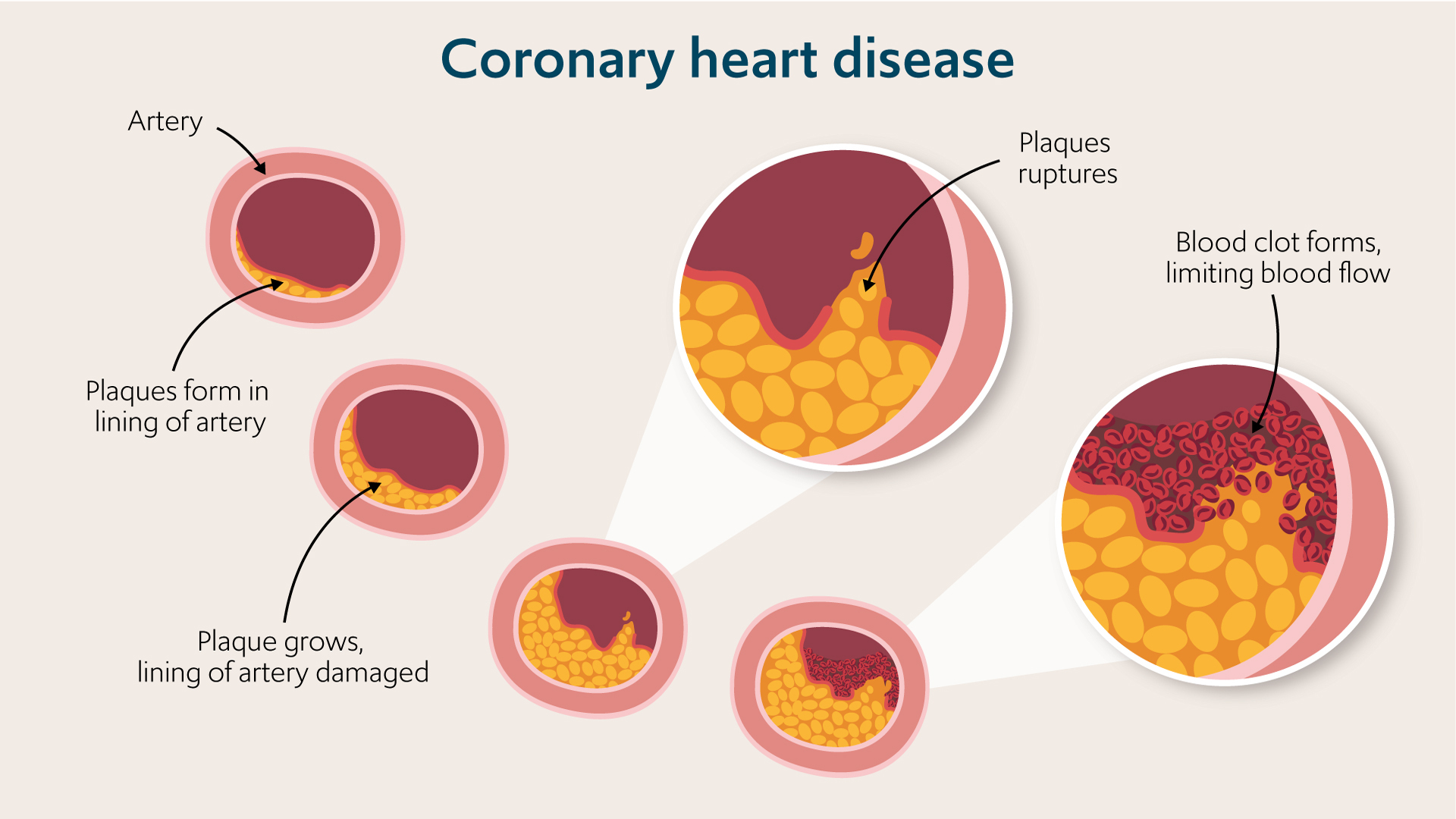
The blood vessels supplying your heart muscles with oxygen are called coronary arteries. The process of plaque build-up in the walls of the coronary arteries leads to the gradual narrowing of blood vessels, and over time, can cause symptoms of physical effort but could remain completely silent before it causes an unexpected heart attack.
It is therefore essential to have a comprehensive assessment of your ongoing symptoms, medical history, and physical examination topped by a range of investigations in order to make a diagnosis of CHD. Early diagnosis of CHD allows prompt and effective treatment to prevent further damage and protect the health of your heart.
“Although many people with coronary heart disease will have the classic symptoms of angina (the medical term for chest pain caused by reduced blood flow to the heart muscle), some patients with significant coronary heart disease may have atypical symptoms or none at all. Therefore for those with significant risk factors medical tests and investigations can help diagnose the presence and magnitude of ischaemia,” explains consultant cardiologist, Dr Aigul Baltabaeva.
How do you diagnose coronary heart disease?
Your GP or cardiologist will take a complete medical history looking at your symptoms, and chronic medical conditions like diabetes and hypertension taking into account other risk factors for coronary heart disease, including age, family history, weight, ethnicity, lifestyle and smoking.
Coronary heart disease symptoms
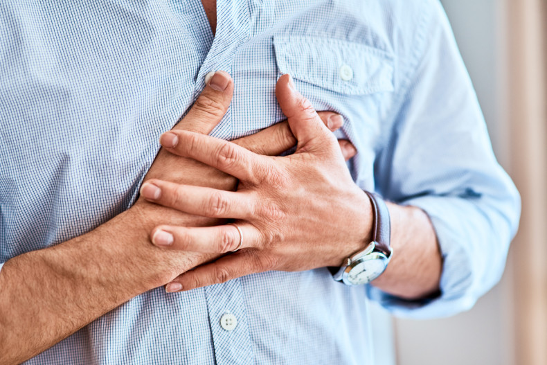
One of the main symptoms of coronary heart disease is chest pain. It may often present as a feeling of dull and heavy discomfort in the middle of the chest or tightness in the chest, sometimes you might feel it as a squeezing or crushing pain under the breastbone. The pain may spread into your arm, jaw, neck or back. Usually, exercise, exertion, cold weather and stress can trigger anginal pain. You may also experience the following symptoms:
- Breathlessness.
- Feeling weak, faint or lightheaded.
- Reeling nauseous and sweaty.
Coronary heart disease examination
Your physical examination may show no evidence of coronary heart disease. However, your doctor will check for several indirect signs and look for other chronic conditions that increase your risk of CHD, such as hypertension and diabetes.
They will also exclude other causes of chest pain, such as lung problems or musculoskeletal pain. The examination may include:
- blood pressure check
- pulse to check for an irregular rhythm
- check for fatty lumps or xanthelasma (yellow growths) on the skin around your eyes and white rings around your irises which may indicate high cholesterol
- examine for evidence of fatty deposits in other blood vessels around the body, which makes it more likely that you also have coronary heart disease. They may check feet pulses, look for swellings in the large blood vessel in the abdomen, the aorta, and listen to the carotid artery in the neck to check if fatty plaques impede the blood supply
- examination for signs of heart failure, including swelling of the feet and ankles and fluid build-up in the lungs
- listen to the heart to check for valve problems
- check your height and weight to calculate BMI.
Depending on your history and examination, your doctor will arrange various tests. The investigation and treatment will depend on whether your doctor assesses that your coronary heart disease is stable or whether you need urgent treatment to protect your health and prevent heart damage. If you have severe chest pain, pain that has come on suddenly or continues when you are resting, you may be having a heart attack that needs emergency medical care.
Investigations for coronary heart disease
Investigations for CHD include blood tests and examinations to assess the structure and function of the heart at rest and during exercise or drug-induced increases in heart rate. Sometimes you might need an assessment of the coronary artery blood flow to help make decisions on the best treatment for your needs.
Blood tests
- Haemoglobin to look for anaemia.
- Glycated haemoglobin to check for diabetes.
- Lipid profile to check the level of cholesterol and ratio of ‘good’ and ‘bad’ cholesterol levels.
- Blood tests to check for thyroid, renal, and liver disease which might contribute to your assessment.
Resting ECG
An electrocardiogram records your heart’s electrical activity which allows your doctor to check how regular and fast your heart beats are. The shape of the curvature of ECG spikes might provide additional information. It is a painless test that takes just a few minutes. The technician places ten sticky patches across your chest and on your limbs, and they connect these through cables to the ECG monitor.
The device picks up and records the electrical activity then prints it on paper. The ECG can identify heart rhythm problems, show if you’re having a heart attack, and indicate if you’ve had one in the past.
Ambulatory ECG recording
You wear an ECG recorder for a prolonged period, often 24 hours but sometimes for much longer up to 2 weeks, as you are going about your everyday life. It can show if your heart remains regular and steady. This test might be needed if you are experiencing palpitations and faintness.
Exercise ECG stress test
Sometimes 12-lead ECG is recorded when exercising on a treadmill or static bike. It shows how ECG changes when you are active. It is not always accurate in detecting changes specific to CHD but is still used in special circumstances.
Chest X-ray
A chest x-ray is an image of your heart, lungs and the bones of your chest wall obtained using a small dose of electromagnetic radiation passing through your chest. It cannot detect CHD but shows whether your heart is enlarged and helps diagnose lung and bone changes.
Coronary CT calcium score and angiography
Coronary CT calcium score and angiography use X-ray images taken rapidly from different angles put together to create images of the heart and blood vessels with special computer processing. CT is short for computerised tomography.
Calcium-score screening tests detect deposits of calcium in the coronary arteries which form with time on atherosclerotic plaques. The high calcium score might indicate a high chance of significant narrowing of blood vessels. If a contrast agent is injected into the veins it is possible to trace it within the blood vessel of the heart and construct 2D and 3D images to locate blockages to the blood flow in your coronary arteries.
“The CT coronary angiogram is less invasive than a traditional angiogram and recommended as a first line imaging technique in a large group of patients with recent onset of chest pain suspected of cardiac origin. It is a safe and relatively quick procedure and allows doctors to provide risk stratification for their patients. It is, however, difficult to know whether moderate coronary narrowing bears functional significance,” explains Dr Baltabaeva.
Echocardiography and stress echo tests
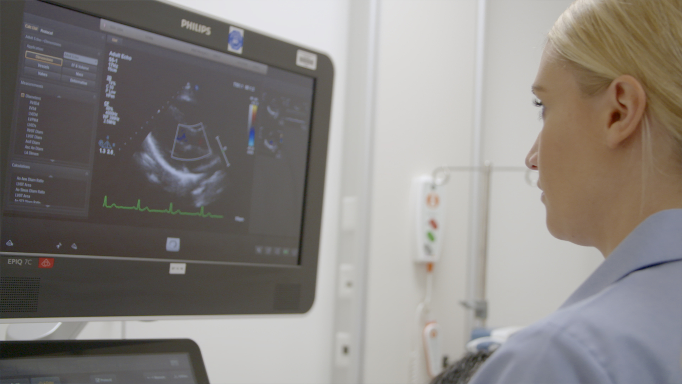
An echocardiogram or ‘echo’ test uses sound waves are sent inside the chest and reflections from heart structures are used to visualise your heart. It is similar to the other ultrasound used in pregnancy or to look at structures in the abdomen and pelvis, but more information is used from echoes of red blood cells to estimate pressures within the heart chambers and the direction of blood flow.
“The moving images show whether all parts of the muscular heart walls are pumping effectively. It is easy to see if the muscle is damaged by a heart attack or coronary heart disease, it becomes thinned and less active than other areas of the heart wall. An echo can also show other heart structures and the valve function. No radiation is used for echo and it can be repeated as many times as needed,” explains Dr Baltabaeva.
Echo images can also be taken during exercise or stimulation of the heart with special medicines making your heart pump stronger and faster. This procedure is called stress echo but there is nothing stressful about this test. Most of the patients tolerate it well without major complications.
If the heart pictures are not clear special dye could be given intravenously to enhance the quality of the images. Stress echo protocol at Harefield Hospital allows us to assess the stiffness of the heart and heart valve function during the procedure.
Cardiac Magnetic resonance (CMR) imaging and stress CMR
CMR uses a powerful magnetic field generated in a large tube-like scanner to create detailed images of your heart. It requires negotiation between the lung and heart movements to acquire clear pictures. You will be asked to hold your breath for a short period of time.
CMR allows us to clearly see the location and size of the scars left behind the heart attacks with the help of a special dye. If you are referred for stress CMR halfway through the test the special agent relaxing the blood vessels will be given through the vein in your arm. The magnet can detect the difference in the blood flow blood vessels fail to relax equally if there has been a build-up of significant plaque. You have to abstain from caffeine for 24 hours prior to the test.
Coronary angiography
Coronary angiography is an invasive investigation that gives detailed information about your heart and coronary arteries. The procedure utilises X-rays and shows images of the coronary arteries taken from multiple angles.
Using special long fine and flexible tubes inserted by the specialist either in your wrist or groin the dye is injected straight into coronary arteries. It shows up in the blood vessels, highlighting any narrowing and blockages. It is possible to dilate the blockage by inflating a small balloon within the blockage and reinforcing it with a special mesh-like tube called ‘stent’ to keep the dilated artery open.
Related content
-
Coronary heart disease
Coronary heart disease is caused by the narrowing of the coronary arteries that supply the heart muscle with oxygen-rich blood.
-
Cardiac catheterisation (coronary angiogram)
Cardiac catheterisation uses x-rays and a dye to take detailed pictures of the arteries.
-
Cardiac MRI scan (CMR)
A cardiac magnetic resonance (CMR) scan is non-invasive and shows detailed images of your heart.